Detecting breast cancer earlier with 3D X-rays
A research team from ETH Zurich and the Paul Scherrer Institute PSI together with the Baden Cantonal Hospital and the University Hospital Zurich wants to improve a method for diagnosing breast cancer.

In brief
- Researchers from the ETH Zurich, the Paul Scherrer Institute PSI, the Cantonal Hospital Baden and the University Hospital Zurich have achieved a milestone on the way to improved breast cancer screening.
- They have demonstrated that breast tumours can be detected earlier with a new type of 3-D X-ray computed tomography than with conventional computed tomography.
- The new grating interferometry computed tomography clearly surpasses the conventional method in terms of resolution and contrast. This will increase the chances of recovery for affected women.
In 2020, breast cancer was the most commonly diagnosed form of cancer worldwide, with over two million cases. In women, it accounts for about a quarter of all cancer cases and is responsible for 15.5 percent of cancer-related deaths. The earlier the diagnosis is confirmed, the sooner the appropriate therapy can begin and the higher the chances of survival. That’s why researchers around the world are working to improve early diagnosis.
And this has now been achieved by a team of researchers from the Paul Scherrer Institute PSI and ETH Zurich together with the Baden Cantonal Hospital (KSB) and the University Hospital Zurich (USZ). They have has succeeded in refining the technique used to detect tumours in their early stages, to produce considerably more reliable results and be less unpleasant for the patients. With this, the researchers have enhanced conventional computed tomography (CT) in a way that significantly improves image resolution without increasing the radiation dose.
This means that small deposits of calcium known as microcalcifications, which indicate breast cancer, could potentially be detected earlier than before. This significantly increases the chances of recovery for affected women. According to the experts, this technique, which is based on X-ray phase contrast, could be swiftly put to use in clinical settings. “We still need a little time,” says Marco Stampanoni, head of the research group at PSI and Professor of X-ray Imaging at ETH Zurich, “but we’ve reached a milestone along the way with our work.”
How effective is mammography?
More accurate early detection of tumours is an important step in the fight against breast cancer. Currently, mammography screening programmes are are used as an early detection tool in many industrialised countries. However, the effectiveness of mammography screening is a controversial topic.
Control studies have found that only 46 percent of suspected cases detected in screening turned out to be actual cases of cancer. A false alarm can cause great distress to sufferers, as it can be two to three weeks before a biopsy result gives the all-clear. Moreover, studies suggest that mammography misses 22 percent of actual cases, lulling those affected into a false sense of security. This is even more serious, as it means valuable time is wasted before treatment can begin.
The weaknesses mentioned here are due to the difficulty that even experts have interpreting mammography images. Soft breast tissue offers only limited contrast on X-ray, while the complicated interior of the breast is often blurred in two-dimensional fluoroscopy. To take X-rays of the breast, it must be squeezed tightly to hold it still. Patients sometimes find this uncomfortable or even painful, which is why some women do not go for screening.
Three-dimensional X-rays bring advantages
In X-ray phase contrast, researchers extend the tumour diagnostics with additional physical information. In other words, the images they create draw on information that conventional X-ray imaging does not take into account: the signals generated when biological tissue refracts and scatters the radiation. When X-rays pass through structures of different densities, they are not only attenuated but also refracted and diffracted. This information can be used to improve both the contrast of the images and their resolution, making the smallest objects easier to identify.
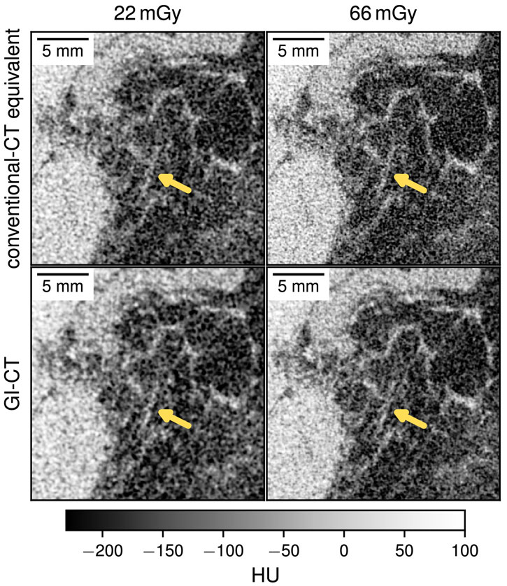
The researchers from PSI, ETH, KSB and USZ use a method familiar from physical metrology called grating interferometry (GI for short). In this, the X-rays pass not only through the object under examination, but also through three gratings with a line spacing of a few micrometres. This makes the additional information visible. In the journal Optica, Stampanoni’s team published several images that clearly demonstrate the advantages of GI computed tomography over conventional X-rays in terms of resolution and contrast.
The X-rays this method requires can be generated with a conventional X-ray source, and the radiation dose is roughly equivalent to that used for conventional computed tomography of the breast. “Our goal is to reduce the dose by a factor of two to three, while maintaining the same resolution, or to increase the resolution by 18 to 45 percent – in each case compared to conventional X-ray,” explains physicist Michał Rawlik, the lead author of the publication and a member of Stampanoni’s research team.
Making cancer screening more comfortable
Assuming they receive authorisation from Swissmedic, Switzerland’s supervisory authority for medical products, the researchers plan to start clinical trials together with USZ and KSB as their clinical partners by the end of 2024. By then, a prototype of the necessary device should be ready to examine patients for the first time. According to Stampanoni, the researchers are expecting the trials will run over a period of one to two years. “If everything goes to plan, we’ll then be able to start developing a commercial device and running studies in selected clinics,” he says.
This new method is also expected to make cancer screening a lot more comfortable. The apparatus will be set up so that the patient rests on her stomach on a couch with cutouts in the chest area. The tomograph itself will be below the couch and shielded from the patient; its measuring device will rotate around the breasts to create a three-dimensional image.
“Phase contrast X-rays make fine details in the tissue visible,” adds Rahel Kubik-Huch, director of the Department of Medical Services at KSB and Chief Physician for Radiology, who was involved in the research. “This translational project will explore the potential to employ this technique in the early detection of breast cancer. KSB is very interested in further advancing the research collaboration with PSI and ETH Zurich. We hope that one day our patients will be able to benefit from these advances.”
This is a slightly edited version of a external pageMedia Release published by the Paul Scherrer Institute PSI. Werner Siefer is a freelance journalist and non-fiction author.
Reference
Rawlik, M et al. Increased dose efficiency of breast CT with grating interferometry. Optica, 18.07.2023. DOI: external page10.1364/OPTICA.487795