Ribosome mapped at high resolution
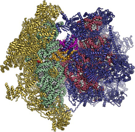
Researchers have succeeded in achieving a high-resolution three-dimensional map of a comparatively large, complex and rare molecule: the complete ribosome (the protein-producing machinery) in the mitochondria (the intracellular powerhouse) of mammals.
Every second, new proteins are produced in every cell of our body as part of its normal metabolism. Ribosomes, molecular complexes composed of numerous components, are the main actors involved in this process. They read the information encoded on a copy of the genetic material and assemble proteins according to this blueprint, building block by building block. Two types of ribosomes are found in the cells of humans and other mammals: the ‘normal’ ribosomes, which occur in large numbers in all cells, and the structurally different and less numerous ribosomes in the mitochondria. These are cellular compartments that are also described as the powerhouses of cells, as they provide the energy for all cellular processes.
The elucidation of the detailed three-dimensional structure of mitochondrial ribosomes is no easy task. On one hand, mitochondrial ribosomes are comparatively large and complex molecules, like all ribosomes. On the other hand, their small numbers make it difficult to obtain highly purified mitochondrial ribosomes. One year ago, ETH researchers led by Nenad Ban, Professor at the Institute of Molecular Biology and Biophysics, and Ruedi Aebersold, Professor at the Institute of Molecular Systems Biology, successfully determined the detailed structure of a subunit of the mitochondrial ribosome of mammals, or, more specifically, of pigs. In further research that has just been published in the journal Science, the same team have now obtained a three-dimensional map of the complete mitochondrial ribosome of pigs.
Molecules ‘in action’
A special feature of the molecular structure now presented is that it shows the ribosome ‘in action’. In addition to the ribosome itself, the ETH researchers were able to map a molecule that contains the blueprint for a protein (termed mRNA) attached to the ribosome. Two tRNA molecules, which deliver protein building blocks to the ribosome, were mapped as well.
The researchers achieved their objective using a combination of two techniques: cryo-electron microscopy, in which a sample is flash frozen before examination, and mass spectrometry. As a result, they were able to obtain the three-dimensional structure of the mammalian mitoribosome with an unprecedented resolution of 3.8 angstroms (less than 0.4 nanometres). “Cryo-electron microscopy has been developed heavily in recent years. Today, we have electron detectors that can correct tiny movements in the sample that would otherwise lead to blurring,” explains ETH professor Ban. The scientists conducted the cryo-electron microscopic experiments at ETH Zurich’s Scientific Center for Optical and Electron Microscopy (ScopeM).
The ribosomes in mitochondria share similarities with those of bacteria. Knowledge of their exact structure is important, amongst others, in pharmaceutical research; for example, in the development of antibiotics that attack the ribosomes in bacteria and keep the micro-organisms in check, but do not lead to any adverse side-effects in human mitochondria.
Literature reference
Greber BJ, Bieri P, Leibundgut M, Leitner A, Aebersold R, Boehringer D, Ban N: The complete structure of the 55S mammalian mitochondrial ribosome. Science, 2 April 2015, doi: external page10.1126/science.aaa3872