How the lungs fight the flu virus
A special type of phagocyte cells in the lungs plays an important role during infection with flu viruses by preserving the exchange of oxygen and carbon dioxide despite massive virus-induced lung tissue damage, as researchers from ETH have demonstrated.
Patients with a serious viral infection caused by the influenza virus often suffer from shortness of breath. Since the virus damages the lung tissue, less oxygen is drawn into the body when the patient inhales. In more serious cases this can even lead to death. Researchers led by Manfred Kopf at the Institute of Molecular Health Sciences at ETH Zurich used mice to analyse how the innate immune system maintains pulmonary functions during viral infections. They found that a specific type of immune cell called the alveolar macrophage plays an important role. “They are absolutely vital for survival,” says Manfred Kopf. In experiments, the researchers infected mice with the H1N1 flu virus. While normal mice were able to survive the infection, those that were genetically altered without alveolar macrophages did not. The reason: their lung function was so severely restricted that they could no longer obtain enough oxygen.
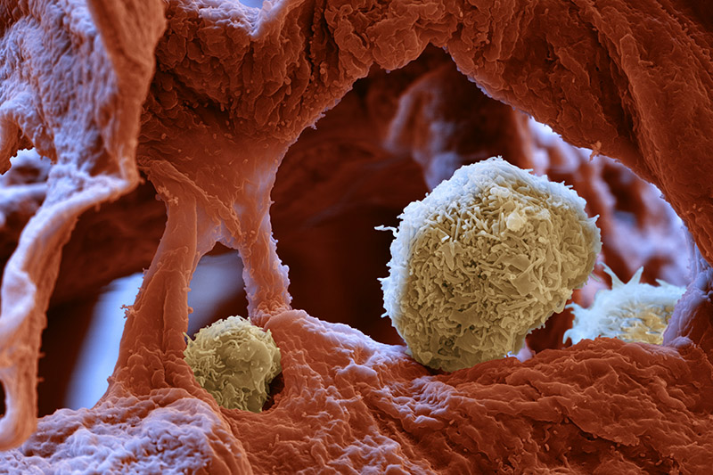
Alveolar macrophages are specialised pulmonary cells found in the pulmonary alveolus. This is where pathogens end up when they are inhaled; they get trapped in the mucus that covers the surface of the pulmonary alveolus. It was previously assumed that the alveolar macrophages primarily protected against bacteria and fungi by consuming the foreign pathogens together with the mucus. “But what we have now found out is that they also protect the organism from the life-threatening complications of a flu infection,” says Kopf.
Cellular vacuum cleaners
Macrophages actually do this indirectly: they do not fight the virus itself, but rather remove debris of the cells that have been destroyed by the virus. “They function like a vacuum cleaner in that they free the lungs from detritus,” says the immunologist. When the phagocyte cells are absent, cellular debris and mucus block the pulmonary alveolus and hinder respiration. This was established in an experiment involving microscopic images of the lungs as well as measurements of blood-oxygen levels. The results of the study were published by the ETH researchers in the journal PLOS Pathogens in April this year.
In a subsequent study, which has recently been published in the journal Nature Immunology, Kopf and his team investigated how the alveolar macrophages come to exist in the first place. Until now it was believed that like other cells in the immune system, they are formed from stem cells in the bone marrow and then migrate through the blood to the pulmonary alveolus throughout a person’s lifetime. By using mouse embryos, the ETH researchers have now been able to establish that this assumption is incorrect. “The progenitors of the alveolar macrophage migrate from the liver into the lungs shortly before birth, where they mature into properly functioning phagocytes after birth,” says Manfred Kopf. The mature cells can then renew themselves in the lungs throughout a person’s life.
Starting point for flu treatments
The researchers were also able to decode which molecules are necessary for this maturation process. Previous studies from other research groups had already indicated that the GM-CSF growth factor plays an important role. This is formed from the epithelial cells in the pulmonary alveolus and acts like a neurotransmitter, binding to the macrophages and activating a signal molecule within them called PPAR-gamma, which controls hundreds of other genes. “That was a surprise,” says Kopf, because before this PPAR-gamma was known primarily for its role in regulating the metabolism of fat cells and insulin sensitivity. Certain medications against adult-onset diabetes, so-called thiazolidinediones, are based on this molecule.
The researchers hope that their findings could one day be used to treat influenza, because activating PPAR-gamma could encourage maturation of the alveolar macrophage and therefore strengthen the immune system. “This could improve a patient’s lung function and help them overcome the illness,” says Kopf. For now, however, this remains speculation as no studies have yet been conducted on human subjects.
Litererature reference
Schneider C, Nobs SP, Kurrer M, Rehrauer H, Thiele C, Kopf M. Induction of the nuclear receptor PPAR-g by the cytokine GM-CSF is critical for the differentiation of fetal monocytes into alveolar macrophages. Nature Immunology, online publication 28 September 2014. doi:external page10.1038/ni.3005
Schneider C, Nobs SP, Heer AK, Kurrer M, Klinke G, van Rooijen N, Vogel J, Kopf M. Alveolar Macrophages Are Essential for Protection from Respiratory Failure and Associated Morbidity following Influenza Virus Infection. PLOS Pathogens, online publication 3 April 2014. doi:external page10.1371/journal.ppat.1004053